Optical Coherence Tomography: Imaging Mouse Retinal Ganglion Cells In Vivo

Scientific Article | Structural changes in the retina are common manifestations of ophthalmic diseases.
Adaptive optics two-photon microscopy enables near-diffraction-limited and functional retinal imaging in vivo
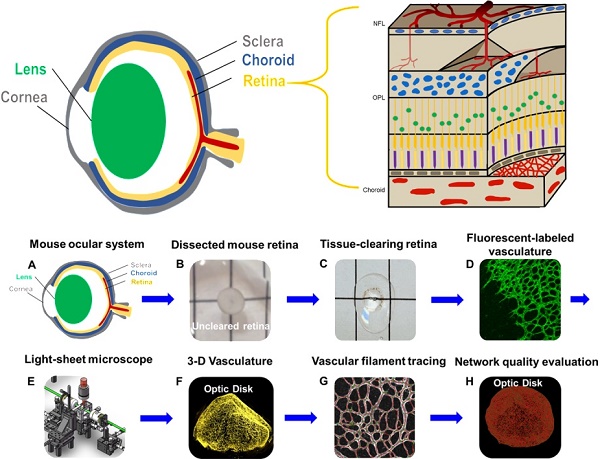
Three-dimensional Imaging Coupled with Topological Quantification Uncovers Retinal Vascular Plexuses Undergoing Obliteration

Emmanuelle SARZI, Professor (Assistant), Claude Bernard University Lyon 1, Villeurbanne, UCBL, Institut NeuroMyogène
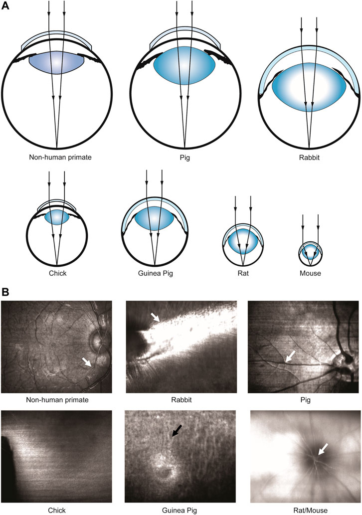
Frontiers In-Vivo Imaging of Ocular Microvasculature Using Swept

Alignment of Visible-Light Optical Coherence Tomography Fibergrams with Confocal Images of the Same Mouse Retina
Frontiers Early Retinal Defects in Fmr1−/y Mice: Toward a Critical Role of Visual Dys-Sensitivity in the Fragile X Syndrome Phenotype?
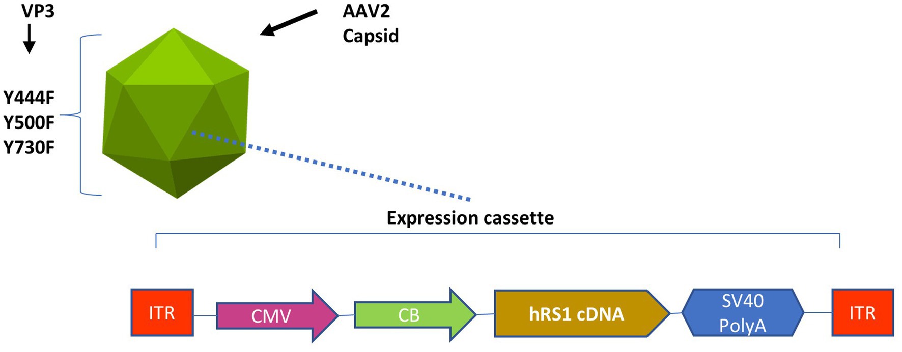
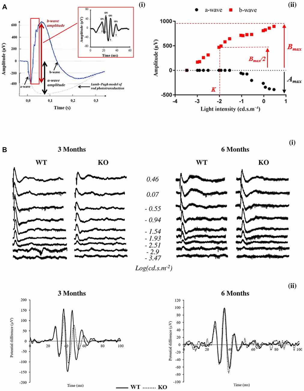
Frontiers The dose-response relationship of subretinal gene therapy with rAAV2tYF-CB-hRS1 in a mouse model of X-linked retinoschisis

Correction-free remotely scanned two-photon in vivo mouse retinal

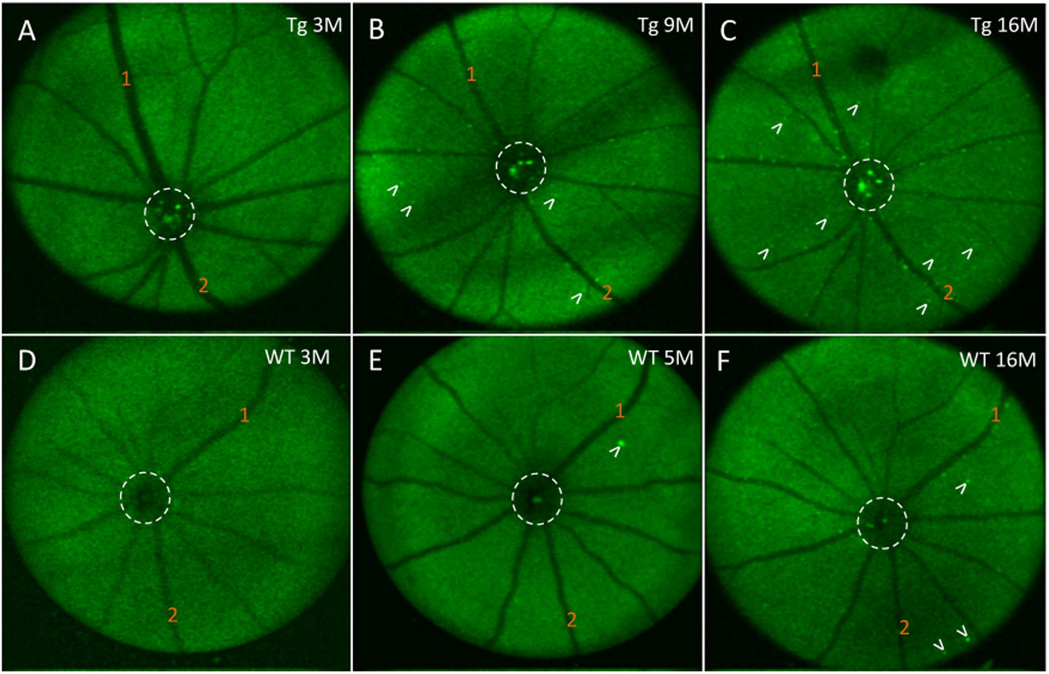
In vivo imaging of adeno-associated viral vector labelled retinal ganglion cells
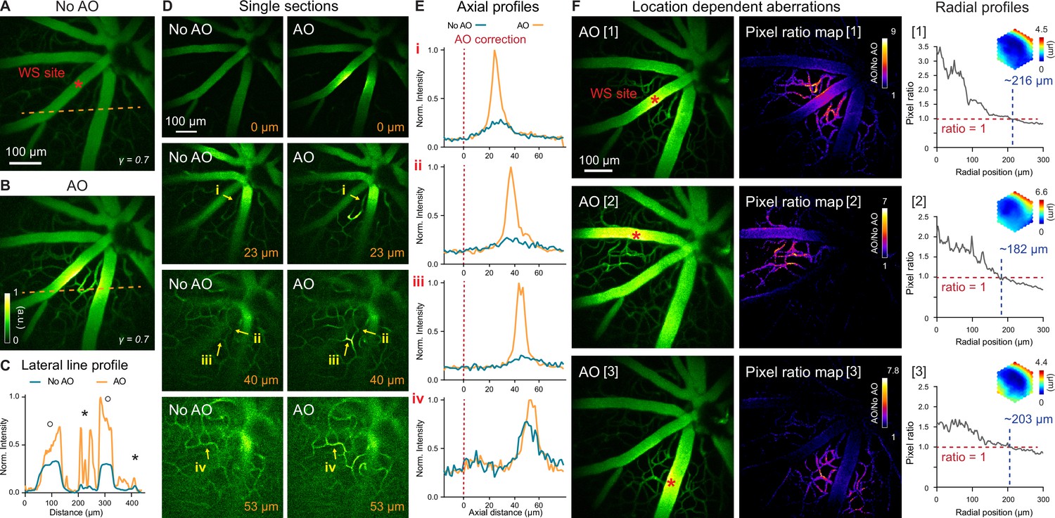
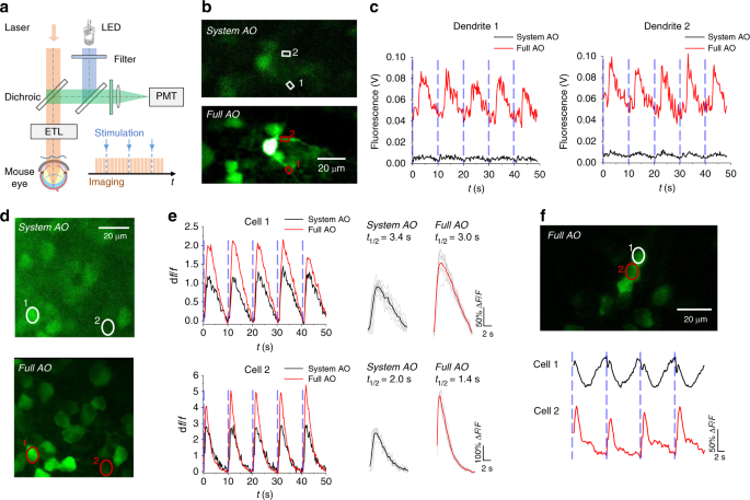
Retinal microvascular and neuronal pathologies probed in vivo by adaptive optical two-photon fluorescence microscopy
Frontiers In vivo Retinal Fluorescence Imaging With Curcumin in an Alzheimer Mouse Model

Fig. 9.4, [In vivo CSLO images of]. - High Resolution Imaging in Microscopy and Ophthalmology - NCBI Bookshelf