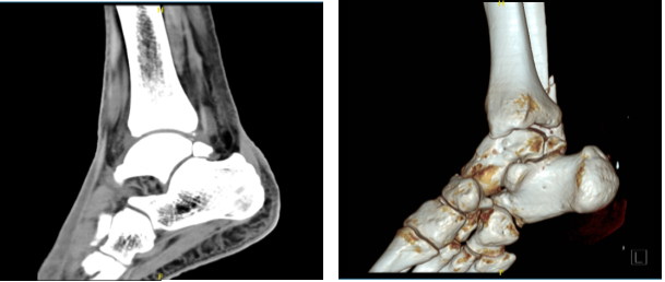
Left Ankle Fracture and Internal Fixation

This exhibit features three radiological colorizations showing an ankle fracture and subsequent internal fixations. The first image depicts a fracture of the distal fibula, fracture of the distal tibia, and disruption of the ankle mortise. The second shows reduction of the fracture fragments with the placement of a fibular plate and multiple screws. Lastly, the third image illustrates fusion of the tibiofibular joint with a syndesmotic screw to reduce widening of the ankle mortise.
Case Study: Fluoroscopic Technique: Open Reduction and Internal
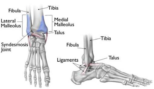
Ankle Fractures (Broken Ankle) - OrthoInfo - AAOS

Immediate continuous passive motion after internal fixation of an

Ankle Fractures - Trauma - Orthobullets

Ankle and Foot Fractures - Physiopedia

This multi-image surgical exhibit features elements associated

ORIF for an Ankle Fracture
Ankle Fracture Boston Medical Center

Open reduction and internal fixation. Mortise view of an unstable

Follow-up conventional X-ray left ankle postoperative after
Left Ankle Fracture and Internal Fixation

JCM, Free Full-Text

Ankle Fractures - Trauma - Orthobullets
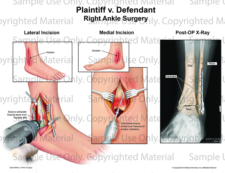
Right Ankle Open Reduction Internal Fixation - MotionLit