Figure 3 from Relevant surgical anatomy of the chest wall.

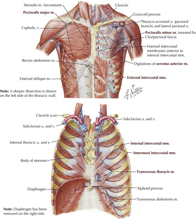
Fig. 3. Anterior chest wall showing the sternum. Note where the costal cartilages articulate with the sternum. In the intercostal space lie different structures: several kinds of intercostal muscles, intercostal arteries and associated veins, lymphatics, and nerves. (From Rendina EA, Ciccone AM. The intercostal space. Thorac Surg Clin 2007;17(4):491e501; with permission.) - "Relevant surgical anatomy of the chest wall."
Thorax Basicmedical Key
Thoracic cavity, Description, Anatomy, & Physiology
Thoracic duct: Anatomy, course and clinical significance
Chest Wall: Anatomy Concise Medical Knowledge

Disorders of the Chest Wall - TeachMeSurgery

Anatomical Variation of the Brachial Plexus and Its Clinical Implications : WFSA - Resources
Lateral abdominal wall reconstruction

SURGICAL ANATOMY OF THE CHEST WALL

Bones and joints of the thoracic wall: Video

Clinical Anatomy & Operative Surgery - Layers of the chest wall. Intercostal space The chest wall can be divided into three layers: a superficial layer of skin and subcutaneous fat; an intermediate

Anatomy of Thoracic Wall.pdf

Trauma nursing 2: management of patients with rib fractures

Thorax - Wikipedia

Anatomical layers of the abdominal and chest walls. A: Surgical